Matsuo, Koichi

写真a

Affiliation

School of Medicine, Collaborative Research Resources (Laboratory of Cell and Tissue Biology) (Shinanomachi)

Position

Professor

Related Websites

External Links

Profile Summary 【 Display / hide

  • Lab of Cell and Tissue Biology (2017)

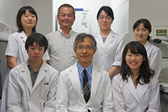
    研究室は、破骨細胞、さらに破骨細胞と骨芽細胞の相互作用(カップリング)に関する一連の骨代謝学の研究で成果を挙げた。近年では、骨の三次元的な形態形成に研究の焦点を移し、内軟骨性骨化や生後の骨形態変化のメカニズムを、マウスやニホンザルなどの実験動物を用いて解析している。「多様なサイズとかたち」を生み出し、それを成長させ、維持し修復する細胞レベルのメカニズムは本質的なものであるにもかかわらず、不明な点が多い。新たな研究領域を開拓しようと、長管骨や椎骨に加え、耳小骨や頭蓋底骨などを対象として解析力を高めている。さらに放射光施設SPring-8(兵庫)におけるX線位相顕微鏡によるCT撮影(東北大学との共同研究)では、 細胞レベルの解像度で、内軟骨性骨化を担う骨形成性毛細血管の構造を明らかにした。研究室を挙げて形態形成原理の追究に精力を注いでいる。

Career 【 Display / hide

  • 2001.04
    -
    2002.03

    Chief, National Institute for Longevity Sciences, Chief

  • 2002.04
    -
    2007.03

    Associate Professor, School of Medicine, Keio University, Department of Microbiology and Immunology, Associate professor

  • 2007.04
    -
    2009.05

    Keio University School of Medicine, Department of Microbiology and Immunology, Associate Professor

  • 2009.06
    -
    Present

    Keio University School of Medicine, Laboratory of Cell and Tissue Biology, Professor

  • 2009.10
    -
    Present

    総合医科学研究センター長

Academic Degrees 【 Display / hide

  • M.D., Ph.D., Keio University, Coursework, 1992.09

Licenses and Qualifications 【 Display / hide

  • 医師免許証, 1986.06


Research Areas 【 Display / hide

  • Bone Metabolism

  • Osteoimmunology

  • Life Science / Cell biology (Cell Biology)

  • Life Science / Anatomy (General Anatomy(includes Histology/Embryology))

Research Keywords 【 Display / hide

  • osteoclast

  • auditory ossicles

  • bone modeling

  • bone remodeling

  • osteocyte

display all >>

Research Themes 【 Display / hide

  • 内軟骨性骨化における骨形成性血管の解析, 

    2015.04
    -
    Present

  • 骨の形態形成とバイオミネラリゼーション・恒常性維持のメカニズムを、細胞間相互作用によって解明することを目指している。, 

    2000
    -
    Present


Papers 【 Display / hide

  • Trans-pairing between osteoclasts and osteoblasts shapes the cranial base during development

    Edamoto M., Kuroda Y., Yoda M., Kawaai K., Matsuo K.

    Scientific Reports (Scientific Reports)  9 ( 1 )  2019.12

     View Summary

    © 2019, The Author(s). Bone growth is linked to expansion of nearby organs, as is the case for the cranial base and the brain. Here, we focused on development of the mouse clivus, a sloping surface of the basioccipital bone, to define mechanisms underlying morphological changes in bone in response to brain enlargement. Histological analysis indicated that both endocranial and ectocranial cortical bone layers in the basioccipital carry the osteoclast surface dorsally and the osteoblast surface ventrally. Finite element analysis of mechanical stress on the clivus revealed that compressive and tensile stresses appeared mainly on respective dorsal and ventral surfaces of the basioccipital bone. Osteoclastic bone resorption occurred primarily in the compression area, whereas areas of bone formation largely coincided with the tension area. These data collectively suggest that compressive and tensile stresses govern respective localization of osteoclasts and osteoblasts. Developmental analysis of the basioccipital bone revealed the clivus to be angled in early postnatal wild-type mice, whereas its slope was less prominent in Tnfsf11 −/− mice, which lack osteoclasts. We propose that osteoclast-osteoblast “trans-pairing” across cortical bone is primarily induced by mechanical stress from growing organs and regulates shape and size of bones that encase the brain.

  • Parathyroid Hormone Shifts Cell Fate of a Leptin Receptor-Marked Stromal Population from Adipogenic to Osteoblastic Lineage

    Yang M., Arai A., Udagawa N., Zhao L., Nishida D., Murakami K., Hiraga T., Takao-Kawabata R., Matsuo K., Komori T., Kobayashi Y., Takahashi N., Isogai Y., Ishizuya T., Yamaguchi A., Mizoguchi T.

    Journal of Bone and Mineral Research (Journal of Bone and Mineral Research)  34 ( 10 ) 1952 - 1963 2019.10

    ISSN  08840431

     View Summary

    © 2019 American Society for Bone and Mineral Research Intermittent parathyroid hormone (iPTH) treatment induces bone anabolic effects that result in the recovery of osteoporotic bone loss. Human PTH is usually given to osteoporotic patients because it induces osteoblastogenesis. However, the mechanism by which PTH stimulates the expansion of stromal cell populations and their maturation toward the osteoblastic cell lineage has not be elucidated. Mouse genetic lineage tracing revealed that iPTH treatment induced osteoblastic differentiation of bone marrow (BM) mesenchymal stem and progenitor cells (MSPCs), which carried the leptin receptor (LepR)-Cre. Although these findings suggested that part of the PTH-induced bone anabolic action is exerted because of osteoblastic commitment of MSPCs, little is known about the in vivo mechanistic details of these processes. Here, we showed that LepR+MSPCs differentiated into type I collagen (Col1)+ mature osteoblasts in response to iPTH treatment. Along with osteoblastogenesis, the number of Col1+ mature osteoblasts increased around the bone surface, although most of them were characterized as quiescent cells. However, the number of LepR-Cre-marked lineage cells in a proliferative state also increased in the vicinity of bone tissue after iPTH treatment. The expression levels of SP7/osterix (Osx) and Col1, which are markers for osteoblasts, were also increased in the LepR+MSPCs population in response to iPTH treatment. In contrast, the expression levels of Cebpb, Pparg, and Zfp467, which are adipocyte markers, decreased in this population. Consistent with these results, iPTH treatment inhibited 5-fluorouracil- or ovariectomy (OVX)-induced LepR+MSPC-derived adipogenesis in BM and increased LepR+MSPC-derived osteoblasts, even under the adipocyte-induced conditions. Treatment of OVX rats with iPTH significantly affected the osteoporotic bone tissue and expansion of the BM adipose tissue. These results indicated that iPTH treatment induced transient proliferation of the LepR+MSPCs and skewed their lineage differentiation from adipocytes toward osteoblasts, resulting in an expanded, quiescent, and mature osteoblast population. © 2019 American Society for Bone and Mineral Research.

  • Innervation of the tibial epiphysis through the intercondylar foramen

    Matsuo K., Ji S., Miya A., Yoda M., Hamada Y., Tanaka T., Takao-Kawabata R., Kawaai K., Kuroda Y., Shibata S.

    Bone (Bone)  120   297 - 304 2019.03

    ISSN  87563282

     View Summary

    © 2018 The Authors The periosteum and mineralized bone are innervated by nerves that sense pain. These include both myelinated and unmyelinated neurons with either free nerve endings or bearing nociceptors. Parasympathetic and sympathetic autonomic nerves also innervate bone. However, little is known about the route sensory nerves take leaving the epiphyses of long bones at the adult knee joint. Here, we used transgenic mice that express fluorescent Venus protein in Schwann cells (Sox10-Venus mice) to visualize myelinated and unmyelinated nerves in the tibial epiphysis. Immunofluorescence to detect a pan-neuronal marker and the sensory neuron markers calcitonin gene-related peptide (CGRP) and tropomyosin receptor kinase A (TrkA) also revealed Schwann cell-associated sensory neurons. Foramina in the intercondylar area of the tibia were conserved between rodents and primates. Venus-labeled fibers were detected within bone marrow of the proximal epiphysis, exited through foramina along with blood vessels in the intercondylar area of the tibia, and joined Venus-labeled fibers of the synovial membrane and meniscus. These data suggest that innervation of the subchondral plate and trabecular bone within the tibial epiphysis carries pain signals from the knee joint to the brain through intercondylar foramina.

  • Bone Marrow Cells Inhibit BMP-2-Induced Osteoblast Activity in the Marrow Environment

    Nguyen H.T., Ono M., Oida Y., Hara E.S., Komori T., Akiyama K., Nguyen H.T.T., Aung K.T., Pham H.T., Tosa I., Takarada T., Matsuo K., Mizoguchi T., Oohashi T., Kuboki T.

    Journal of Bone and Mineral Research (Journal of Bone and Mineral Research)  34 ( 2 ) 327 - 332 2019.02

    ISSN  08840431

     View Summary

    © 2018 American Society for Bone and Mineral Research Bone morphogenetic protein 2 (BMP-2) is widely known as a potent growth factor that promotes bone formation. However, an increasing number of studies have demonstrated side effects of BMP-2 therapy. A deeper understanding of the effect of BMP-2 on cells other than those involved directly in bone remodeling is of fundamental importance to promote a more effective delivery of BMP-2 to patients. In this study, we aimed to investigate the effect of BMP-2 in the marrow environment. First, BMP-2 adsorbed onto titanium implants was delivered at the tooth extraction socket (marrow-absent site) or in the mandible marrow of beagle dogs. BMP-2 could induce marked bone formation around the implant at the tooth extraction socket. Surprisingly, however, no bone formation was observed in the BMP-2-coated titanium implants inserted in the mandible marrow. In C57BL/6 mice, BMP-2 adsorbed in freeze-dried collagen pellets could induce bone formation in marrow-absent calvarial bone. However, similar to the canine model, BMP-2 could not induce bone formation in the femur marrow. Analysis of osteoblast differentiation using Col1a1(2.3)-GFP transgenic mice revealed a scarce number of osteoblasts in BMP-2-treated femurs, whereas in the control group, osteoblasts were abundant. Ablation of femur marrow recovered the BMP-2 ability to induce bone formation. In vitro experiments analyzing luciferase activity of C2C12 cells with the BMP-responsive element and alkaline phosphatase activity of MC3T3-E1 osteoblasts further revealed that bone marrow cells inhibit the BMP-2 effect on osteoblasts by direct cell–cell contact. Collectively, these results showed that the effect of BMP-2 in inducing bone formation is remarkably repressed by marrow cells via direct cell–cell contact with osteoblasts; this opens new perspectives on the clarification of the side-effects associated with BMP-2 application. © 2018 American Society for Bone and Mineral Research.

  • Effects of long-term cigarette smoke exposure on bone metabolism, structure, and quality in a mouse model of emphysema

    Sasaki Mamoru, Chubachi Shotaro, Kameyama Naofumi, Sato Minako, Haraguchi Mizuha, Miyazaki Masaki, Takahashi Saeko, Nakano Takayoshi, Kuroda Yukiko, Betsuyaku Tomoko, Matsuo Koichi

    PLoS One (PLoS ONE)  13 ( 1 )  2018.01

    ISSN  1932-6203

     View Summary

    <p>Smoking is a common risk factor for both chronic obstructive pulmonary disease (COPD) and osteoporosis. In patients with COPD, severe emphysema is a risk factor for vertebral fracture; however, the effects of smoking or emphysema on bone health remain largely unknown. We report bone deterioration in a mouse model of emphysema induced by nose-only cigarette smoke (CS) exposure. Unexpectedly, short-term exposure for 4-weeks decreased bone turnover and increased bone volume in mice. However, prolonged exposure for 20- and 40-weeks reversed the effects from suppression to promotion of bone resorption. This long-term CS exposure increased osteoclast number and impaired bone growth, while it increased bone volume. Strikingly, long-term CS exposure deteriorated bone quality of the lumbar vertebrae as illustrated by disorientation of collagen fibers and the biological apatite c-axis. This animal model may provide a better understanding of the mechanisms underlying the deterioration of bone quality in pulmonary emphysema caused by smoking.</p>

display all >>

Papers, etc., Registered in KOARA 【 Display / hide

display all >>

Reviews, Commentaries, etc. 【 Display / hide

  • Bone Cell Biology Assessed by Microscopic Approach. Regulation of bone mineralization through the osteocyte lacuno-canalicular network

    Matsuo Koichi

    Clinical calcium 25 ( 10 ) 1461 - 1466 2015.10

    Article, review, commentary, editorial, etc. (trade magazine, newspaper, online media), Single Work,  ISSN  0917-5857

     View Summary

    <p>Osteocytes and their dendrites form a large network called the lacuno-canalicular system in mammalian bone. Osteocytes are believed to directly contribute to regulation of mineralization and demineralization in bone matrix, in addition to their indirect regulation of these processes through osteoblasts and osteoclasts. Not only can the location and shape of osteocyte lacunae and canaliculi be spatially visualized in isolated bone samples using synchrotron radiation technology, but differences in the degree of mineralization throughout the lacuno-canalicular system can be detected and quantified. Currently, comparable observation of the time course of these activities in vivo is technically challenging. This review provides an overview of non-dynamic quantitative analysis in the lacuno-canalicular system. Such analysis has the potential to become a methodological basis for investigating osteocyte-dependent direct regulation of mineralization in bone diseases. </p>

  • [Glucocorticoid and Bone. Osteocytic osteolysis:potential modulation by glucocorticoids.

    Matsuo Koichi

    Clinical calcium 24 ( 9 ) 1337 - 1342 2014.09

    Article, review, commentary, editorial, etc. (trade magazine, newspaper, online media), Single Work,  ISSN  0917-5857

  • [Osteoimmunologic regulation of aging].

    Matsuo Koichi

    Clinical calcium 23 ( 1 ) 59 - 64 2013.01

    Article, review, commentary, editorial, etc. (trade magazine, newspaper, online media), Single Work,  ISSN  0917-5857

     View Summary

    <p>In bones, aging manifests itself as a shift towards production of myeloid cells in bone marrow, a condition associated with increased chronic inflammation by macrophages and decreased bone mass due to excess bone resorption by osteoclasts. An increase in the ratio of RANKL, a cytokine promoting osteoclast differentiation, to osteoprotegerin (OPG) , which acts as a decoy RANKL receptor, cannot explain the observed increase in osteoclast production, as serum OPG levels increase with age in humans, apparently to levels insufficient to counteract bone loss and prevent fracture. Age-related increases in osteoclastogenesis, decreases in lymphopoiesis, and inflammation including arthritis are likely best explained by a vicious cycle of myeloid skewing and inflammation occurring in bone marrow. These activities are due to aging of both hematopoietic stem cells themselves and the bone marrow microenvironment (niche cells) , which supports hematopoiesis. Impaired osteoblastogenesis and niche cell function are most likely pathologies emerging from increased oxidative stress, peroxisome proliferator-activated receptorγ (PPARγ) activity, and adipogenesis in the aging bone marrow. Currently, administration of either OPG or an anti-RANKL antibody has proved beneficial to prevent block bone dysfunction and osteoporosis in the elderly. However, anti-aging interventions targeting mesenchymal stem cell differentiation in the bone marrow may also help counteract inflammation and osteoclastic bone loss and enhance osteoblastic bone formation.</p>

  • Regulation of bone metabolism by Eph-ephrin family members

    Matsuo K、, Otaki N

    Clinical calcium 22 ( 11 ) 1669 - 1675 2012.11

    Article, review, commentary, editorial, etc. (trade magazine, newspaper, online media), Joint Work,  ISSN  0917-5857

  • [Osteocytic osteolysis

    Matsuo Koichi, Nango Nobuhito

    Clinical calcium 22 ( 5 ) 677 - 683 2012.05

    Article, review, commentary, editorial, etc. (trade magazine, newspaper, online media),  ISSN  0917-5857

     View Summary

    <p>Osteocytes are present in osteocytic lacunae in both cortical and trabecular bone, where they are interconnected by numerous dendrites in osteocytic canaliculi. In mammals, osteocytes are the most abundant bone cells, outnumbering osteoclasts and osteoblasts. Osteoclasts are the primary bone-resorbing cells ; however, the concept that osteocytes resorb bone by a process called osteocytic osteolysis has been postulated to explain dynamic calcium release from bone in conditions as diverse as parathyroid hormone (PTH) stimulation, hibernation, glucocorticoid stimulation, and lactation. Osteocytic osteolysis remains a controversial concept, mainly because it is difficult to demonstrate experimentally. Recently, novel functions of osteocytes in mineral metabolism and bone remodeling have been reported, and osteocytic osteolysis is being examined more closely experimentally. This review discusses published literature relevant to osteocytic osteolysis and compares 2D and 3D measurements of the volume of osteocytic lacunae, which serve as anatomical evidence for "periosteocytic osteolysis" .</p>

display all >>

Presentations 【 Display / hide

  • Mammalian-type Brn-1/Pou3f3 with 'homopolymeric amino acids (HPAAs)' is essential for stress-induced bone metabolic changes in mice

    Igarashi Atsushi, Matsuo Koichi, Ueda Shintaroh

    Australian New Zealand Bone & Mineral Society Annual Scientific Meeting (Hobart, Tasmania) , 

    2015.11

    Poster presentation

  • Seasonality in Bone Mineralization of Auditory Ossicles and Long Bones in the Primate Macaca fuscata

    Matsuo Koichi

    Annual Meeting of the American Society for Bone and Mineral Research (Seattle, Washington, USA) , 

    2015.10

    Poster presentation

  • Sialylated Glycans of MMP-9 Marak Bone Resorption Lacunae.

    Kuroda Yukiko, Kuno Atsushi, Narimatsu Hisashi,Matsuo Koichi

    Annual Meeting of the American Society for Bone and Mineral Research (Seattle, Washington, USA) , 

    2015.10

    Poster presentation

  • Continuous Parathyroid Hormone Injection in Mouse Has Differential Effects on Osteoclast Activation in Primary and Secondary Spongiosa.

    Nango Nobuhito, Kubota Shogo, Yashiro Wataru, Momose Atsushi, Ichinose Shizuko,Matsuo Koichi

    Annual Meeting of the American Society for Bone and Mineral Research (Seattle, Washington, USA) , 

    2015.10

    Poster presentation

  • EphB/ephrin-B interactions regulate stromal cell fate determination and bone marrow support

    Matsuo Koichi

    Annual Meeting of the American Society for Bone and Mineral Research (Seattle, Washington, USA) , 

    2015.10

    Poster presentation

display all >>

Research Projects of Competitive Funds, etc. 【 Display / hide

  • Construction technique for a bilateral skeleton composed of homochiral building blocks

    2021.09
    -
    2023.03

    MEXT,JSPS, Grant-in-Aid for Scientific Research, 学術変革領域研究(A), Principal investigator

  • Understanding the cellular basis of left-right symmetry in the skeleton

    2021.04
    -
    2025.03

    MEXT,JSPS, Grant-in-Aid for Scientific Research, Grant-in-Aid for Scientific Research (B), Principal investigator

  • Osteogenic capillaries - new aspect of endochondral ossification

    2017.04
    -
    2021.03

    MEXT,JSPS, Grant-in-Aid for Scientific Research, Grant-in-Aid for Scientific Research (B), Principal investigator

Awards 【 Display / hide

  • Distinguished Scientist Award

    2014.07, Japanese Society for Bone and Mineral Research

  • Kitasato Award

    2003.06, Keio University School of Medicine

  • Young Investigator Award

    1997.09, American Society for Bone and Mineral Research


Courses Taught 【 Display / hide

  • MOLECULAR CELL BIOLOGY

    2024

  • MICROBIOLOGY

    2024

  • MEDICAL PROFESSIONALISM 3

    2024

  • INTRODUCTION TO MEDICINE

    2024

  • INTERNSHIP

    2024

display all >>

Courses Previously Taught 【 Display / hide

  • Molecular Cell Biology (MCB) II

    Keio University

    2015.04
    -
    2016.03

    Spring Semester, Lecture

  • Microbiology

    Keio University

    2015.04
    -
    2016.03

    Spring Semester, Lecture

  • Medical Professionalism III (Research Ethics)

    Keio University

    2015.04
    -
    2016.03

    Spring Semester, Lecture

  • Molecular Cell Biology (MCB) II

    Keio University

    2014.04
    -
    2015.03

    Spring Semester, Lecture

  • Microbiology

    Keio University

    2014.04
    -
    2015.03

    Spring Semester, Lecture

display all >>


Memberships in Academic Societies 【 Display / hide

  • The Japanese Society for Bone and Mineral Research

     
  • American Society for Bone and Mineral Research (ASBMR)

     
  • International Bone and Mineral Metabolism (IBMS)